

NEFL Participates in the Progression of Alzheimer's Disease through the TSC1-mTOR Signaling Pathway
-
摘要:
目的 探讨NEFL(neurofilament light chain)通过与TSC1(tuberous sclerosis complex 1)相互作用影响阿尔兹海默病(Alzheimer’s disease,AD)进展的潜在机制。 方法 成年雄性SD大鼠构建AD模型,物体识别测试和转棒实验评估各组大鼠的认知和运动功能。β淀粉样蛋白1-42(Amyloid β Peptide 1-42,Aβ1-42)处理神经元细胞共用体建外细胞模型。新物体识别测试和转棒实验评估各组大鼠的认知和运动功能。RT-qPCR检测NEFL和TSC1的表达。Western blot检测NEFL、TSC1、p-TSC1、mTOR、p-mTOR、S6K1、p-S6K1蛋白的表达水平。CCK-8试剂盒检测细胞活力。ELISA试剂盒检测细胞和组织中IL-6、IL-1β、TNF-α、Aβ1-42、p-Tau的含量。生化试剂盒检测细胞和组织中GSH、MDA、SOD、AChE的含量。流式细胞术检测细胞凋亡率。免疫共沉淀实验检测NEFL和TSC1的相互作用。 结果 成功建立了大鼠AD模型。NEFL在AD大鼠海马组织中表达显著上调,TSC1显著下调。敲低NEFL可减轻AD大鼠的认知和运动功能障碍,降低AD大鼠海马组织中炎症反应,减少氧化应激损伤,并抑制p-mTOR和p-S6K1蛋白表达(P < 0.05)。在Aβ1-42诱导的神经元中,NEFL表达显著升高(P < 0.0001 ),TSC1表达显著降低(P <0.0001 );敲低NEFL后TSC1表达显著升高(P <0.0001 ),细胞凋亡率显著降低(P <0.0001 ),细胞活力显著恢复(P <0.0001 )。敲低NEFL还可降低Aβ1-42诱导的IL-1β、IL-6和TNF-α水平(P <0.0001 ),抑制MDA和AChE水平升高(P < 0.001),并提高GSH和SOD水平(P < 0.01)。与sh-NC组相比,sh-NEFL组p-mTOR和p-S6K1蛋白表达显著下调(P < 0.001)。Co-IP结果显示NEFL与TSC1存在相互作用;敲低NEFL可上调TSC1和p-TSC1蛋白表达(P < 0.01),而过表达TSC1可抑制NEFL蛋白表达(P < 0.05)。敲低TSC1能够逆转敲低NEFL的作用效果。结论 敲低NEFL不仅能够减轻炎症反应,改善氧化应激状态,还可能通过抑制mTOR/S6K1信号通路发挥神经保护作用。 Abstract:Objective To investigate the potential mechanism by which neurofilament light chain (NEFL) affects the progression of Alzheimer’ s disease (AD) through its interaction with tuberous sclerosis complex 1 (TSC1). Methods An AD model was established in adult male Sprague-Dawley rats. Cognitive and motor functions were evaluated using the novel object recognition test and the rotarod test. An in vitro cell model was established by treating neuronal cells with amyloid β peptide 1–42 (Aβ1–42). RT-qPCR was used to detect the expression of NEFL and TSC1. Western blot was used to detect the expression levels of NEFL, TSC1, p-TSC1, mTOR, p-mTOR, S6K1, and p-S6K1 proteins. The CCK-8 kit was used to detect cell viability. ELISA kit was used to detect the contents of IL-6, IL-1β, TNF-α, Aβ1-42, and p-Tau in cells and tissues. Biochemical kits were used to detect the levels of GSH, MDA, SOD, and AChE in cells and tissues. Flow cytometry was used to detect the apoptosis rates. Co-immunoprecipitation assay was used to detect the interaction between NEFL and TSC1. Results An AD rat model was successfully established. The expression of NEFL was significantly upregulated in the hippocampal tissue of AD rats, while TSC1 expression was significantly downregulated. Knockdown of NEFL alleviated cognitive and motor dysfunction in AD rats, reduced inflammatory responses and oxidative stress injury in hippocampal tissues, and inhibited the protein expression of p-mTOR and p-S6K1 (P < 0.05). In Aβ1-42-induced neurons, NEFL expression was significantly increased (P < 0.0001 ), while TSC1 expression was significantly decreased (P <0.0001 ). After NEFL knockdown, TSC1 expression was significantly increased (P <0.0001 ), cell apoptosis rate was significantly reduced (P <0.0001 ), and cell viability was significantly restored (P <0.0001 ). NEFL knockdown also decreased the levels of IL-1β, IL-6, and TNF-α induced by Aβ1-42 (P <0.0001 ), suppressed the elevation of MDA and AChE (P <0.0001 ), and increased the levels of GSH and SOD (P < 0.01). Compared with the sh-NC group, the protein expression levels of p-mTOR and p-S6K1 (P < 0.001) were significantly downregulated in the sh-NEFL group. Co-immunoprecipitation results showed an interaction between NEFL and TSC1. Knockdown of NEFL upregulated the protein expression of TSC1 and p-TSC1 (P < 0.01), whereas overexpression of TSC1 inhibited NEFL protein expression (P < 0.05). Moreover, knockdown of TSC1 reversed the effects of NEFL knockdown.Conclusion Knockdown of NEFL not only alleviates inflammatory response and improves oxidative stress status, but may also exert neuroprotective effects by inhibiting the mTOR/S6K1 signaling pathway. -
Key words:
- Alzheimer’ s disease /
- NEFL /
- TSC1 /
- Neuroinflammation /
- Oxidative stress
-
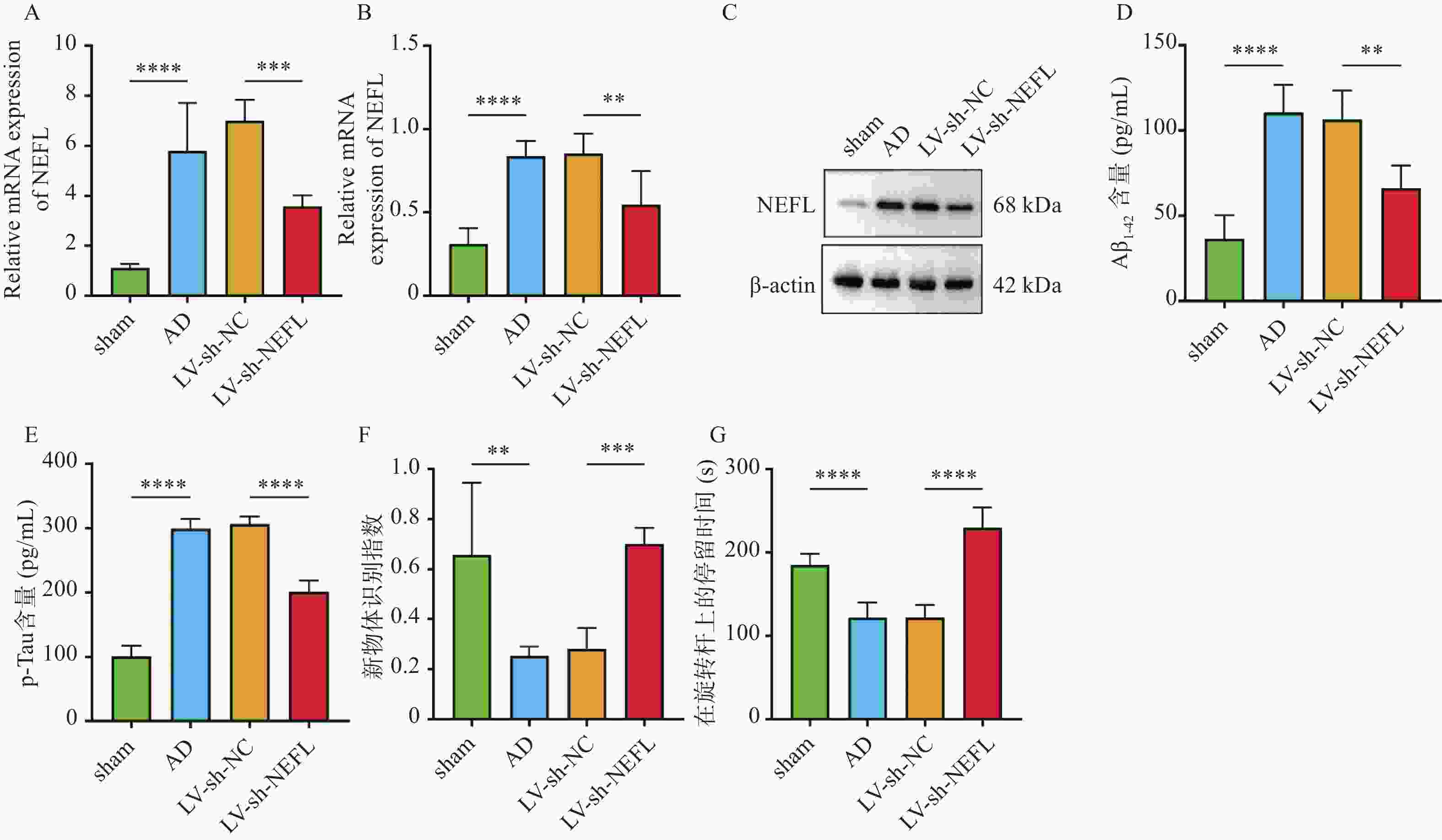
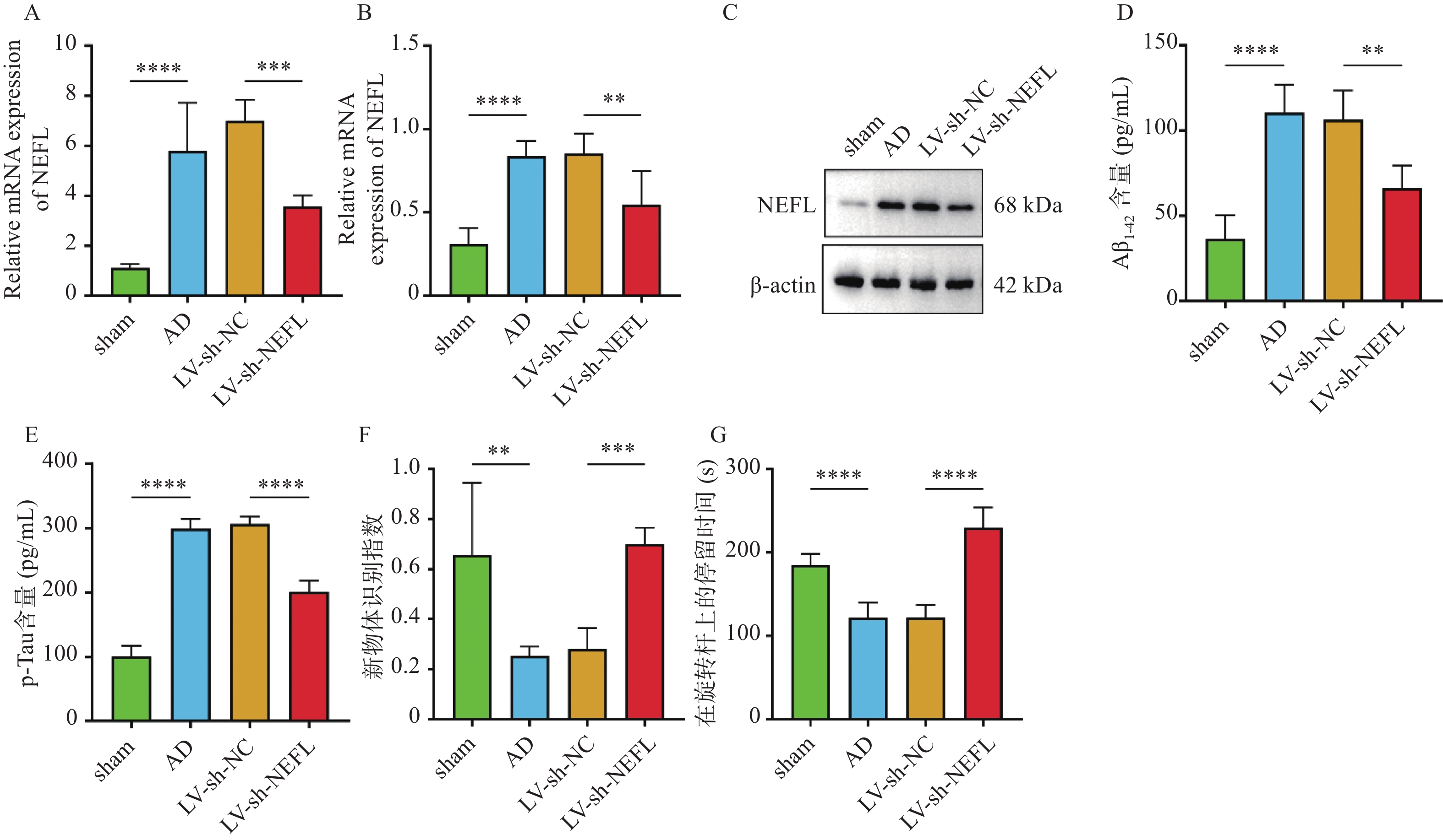
图 1 NEFL对AD大鼠的影响 ($ \bar x \pm s $,n = 6)
A:RT-qPCR检测NEFL的表达;B~C:Western blot检测NEFL的表达;D:ELISA试剂盒检测Aβ1-42的含量;E:ELISA试剂盒检测p-Tau的含量;F:新物体识别指数;*P < 0.05;**P < 0.01;****P < 0.0001。
Figure 1. Effects of NEFL on AD rats ($ \bar x \pm s $,n = 6)

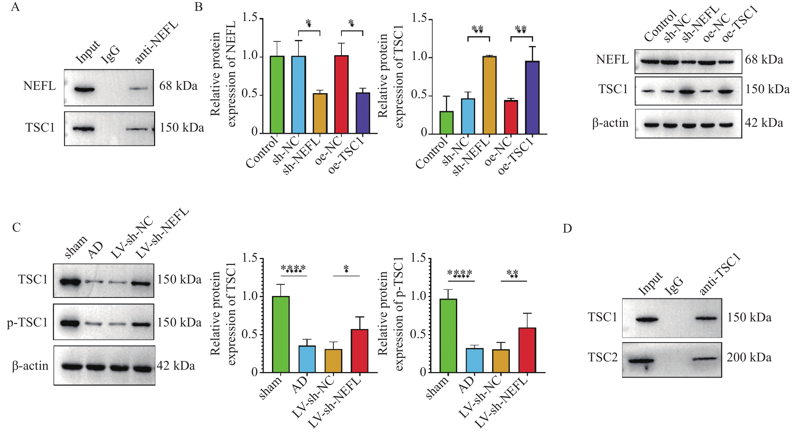
图 5 NEFL与TSC1相互作用 ($ \bar x \pm s $,n = 3)
A:Co-IP实验检测NEFL与TSC1的相互作用;B:Western blot检测NEFL和TSC1蛋白表达;C:Western blot检测TSC1和p-TSC1蛋白表达;D:Co-IP实验检测TSC1与TSC2的相互作用;**P < 0.01;***P < 0.001;****P < 0.0001。
Figure 5. Interaction between NEFL and TSC1 ($ \bar x \pm s $,n = 3)
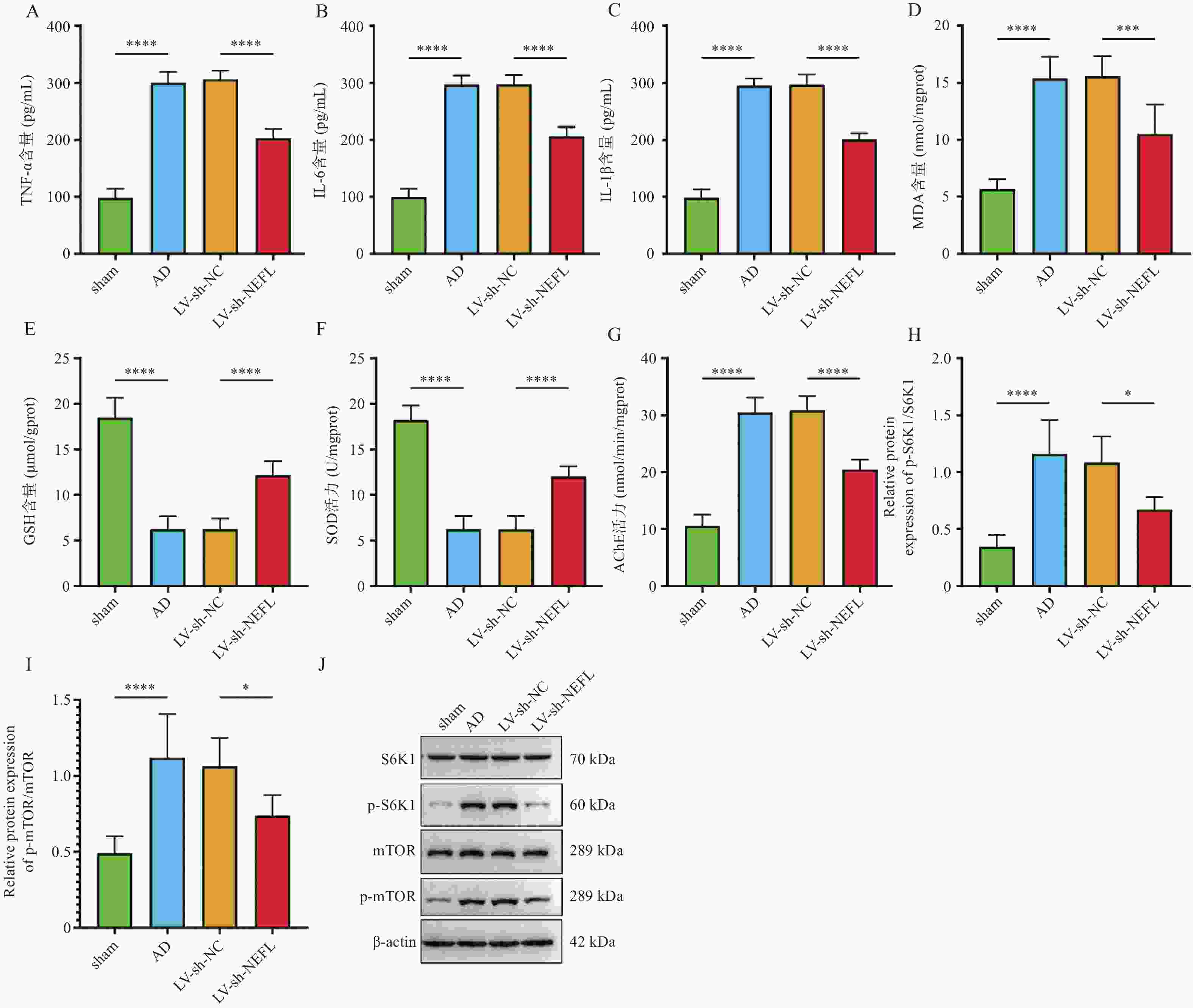
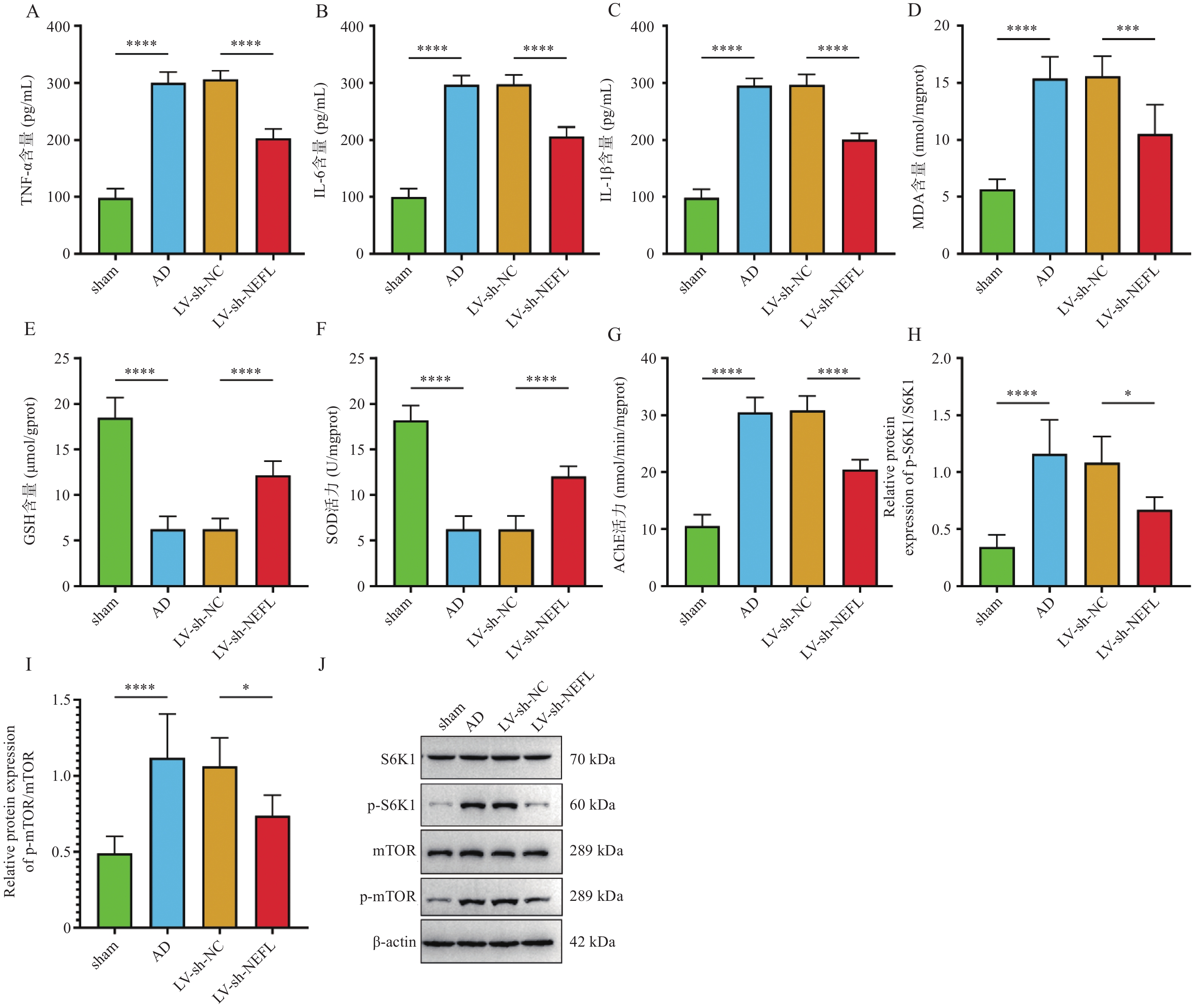
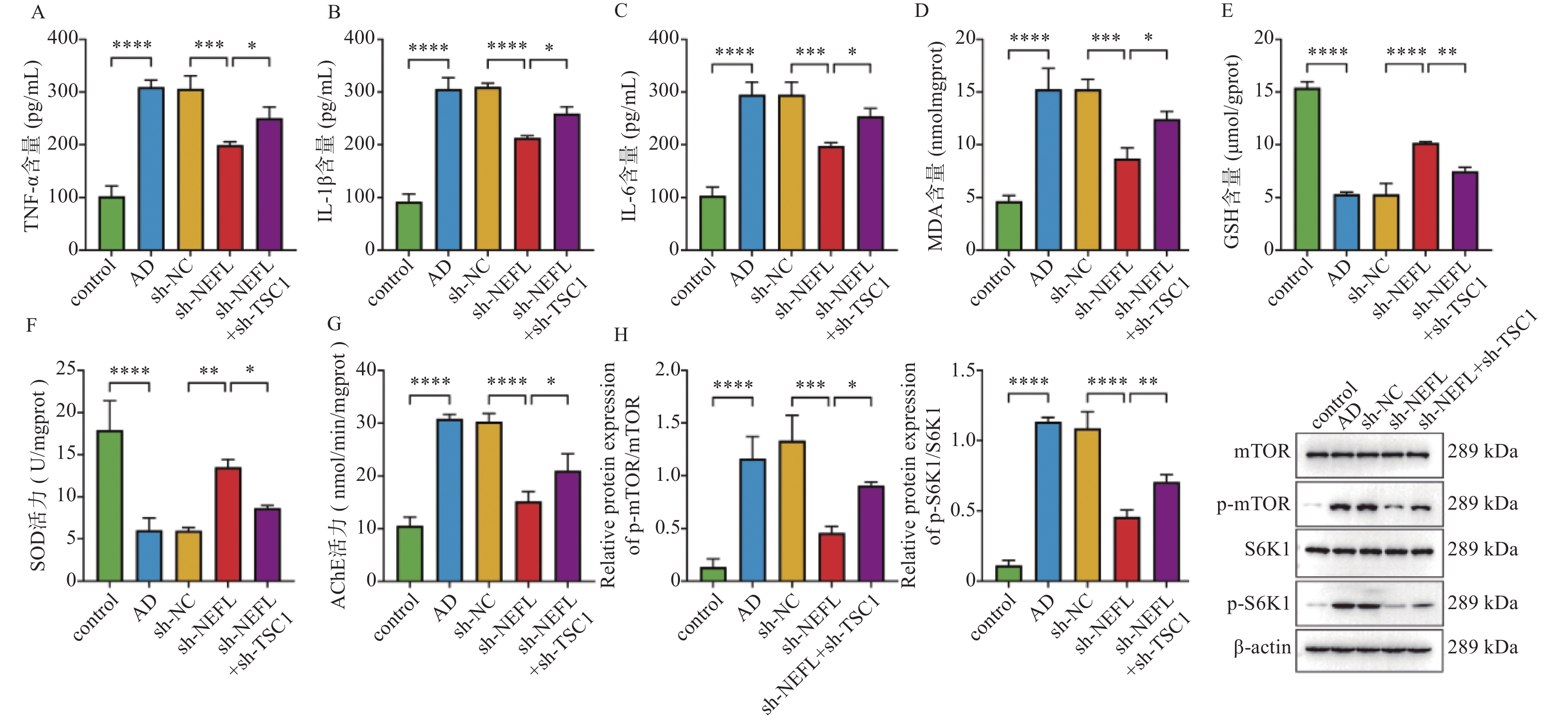
图 2 敲低NEFL对AD大鼠的影响 ($ \bar x \pm s $,n = 6)
A~C:ELISA检测TNF-α、IL-1β、IL-6的含量;D~G:生化检测试剂盒检测MDA、GSH、SOD、AChE的含量;H~J:Western blot检测mTOR/S6K1通路相关蛋白的表达;**P < 0.01;***P < 0.001;****P < 0.0001。
Figure 2. Effects of NEFL knockdown on AD rats ($ \bar x \pm s $,n = 6)
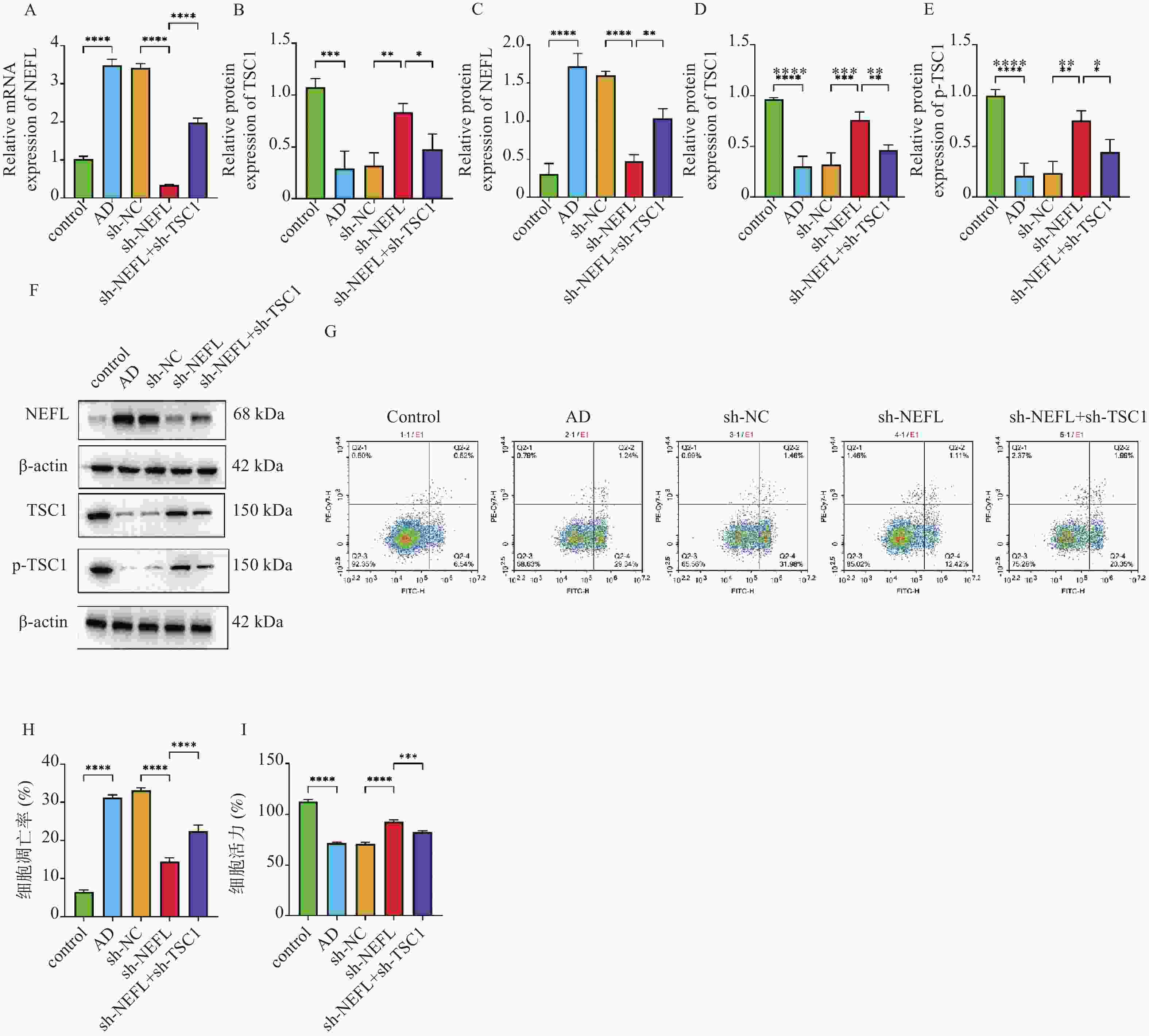
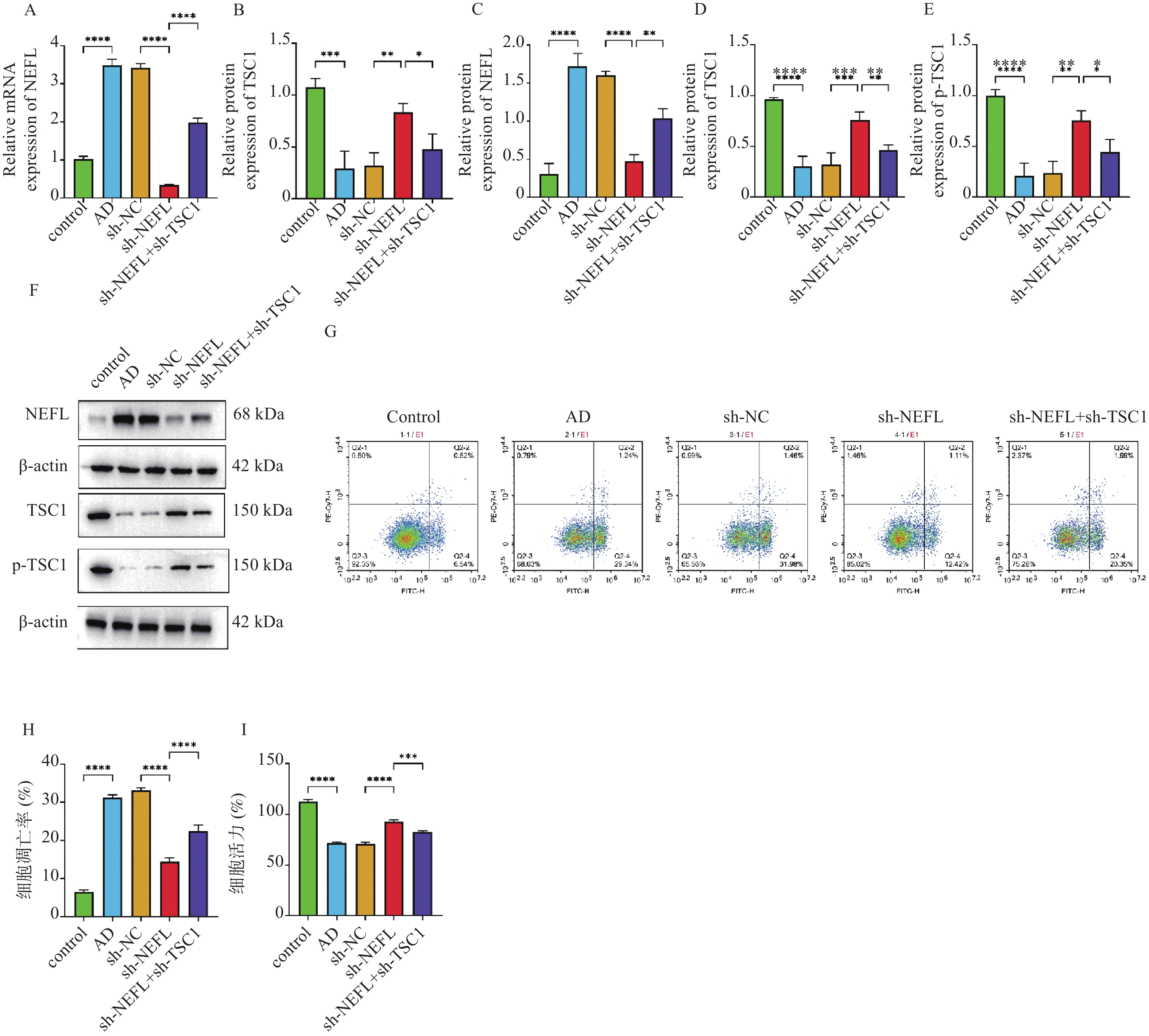
图 3 NEFL对Aβ1-42诱导的细胞损伤的影响 ($ \bar x \pm s $,n = 3)
A~B:RT-qPCR检测NEFL和TSC1的表达;C~F:Western blot检测NEFL、TSC1、p-TSC1的表达;G~H:流式细胞术检测细胞凋亡;I:CCK-8检测细胞活力;**P < 0.01;***P < 0.001;****P < 0.0001。
Figure 3. Effects of NEFL on Aβ1-42-induced cellular damage ($ \bar x \pm s $,n = 3)
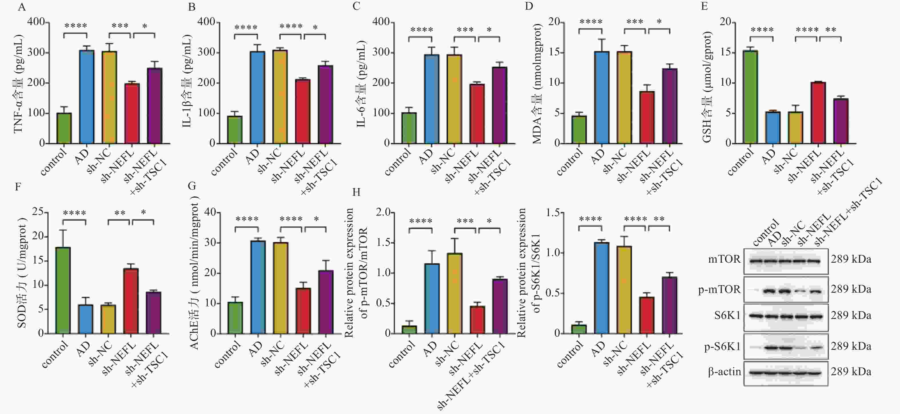
图 4 NEFL对Aβ1-42诱导的细胞炎症和mTOR/S6K1通路的影响 ($ \bar x \pm s $,n = 3)
A~C:ELISA检测TNF-α、IL-1β、IL-6的含量;D~G:生化检测试剂盒检测MDA、GSH、SOD、AChE的含量;H:Western blot检测mTOR/S6K1通路相关蛋白的表达;*P < 0.05;**P < 0.01;***P < 0.001;****P < 0.0001。
Figure 4. Effects of NEFL on Aβ1-42-induced cellular inflammation and the mTOR/S6K1 pathway ($ \bar x \pm s $,n = 3)
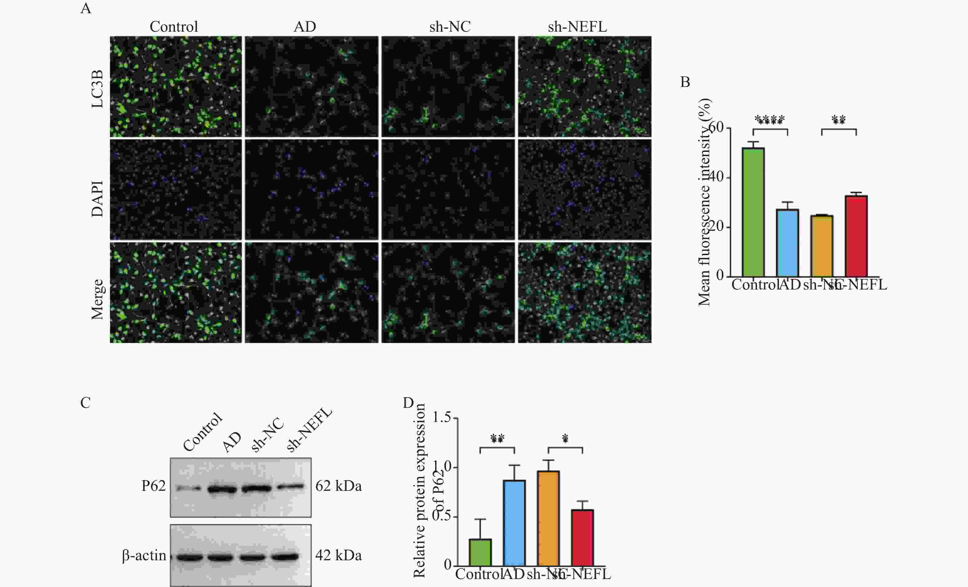
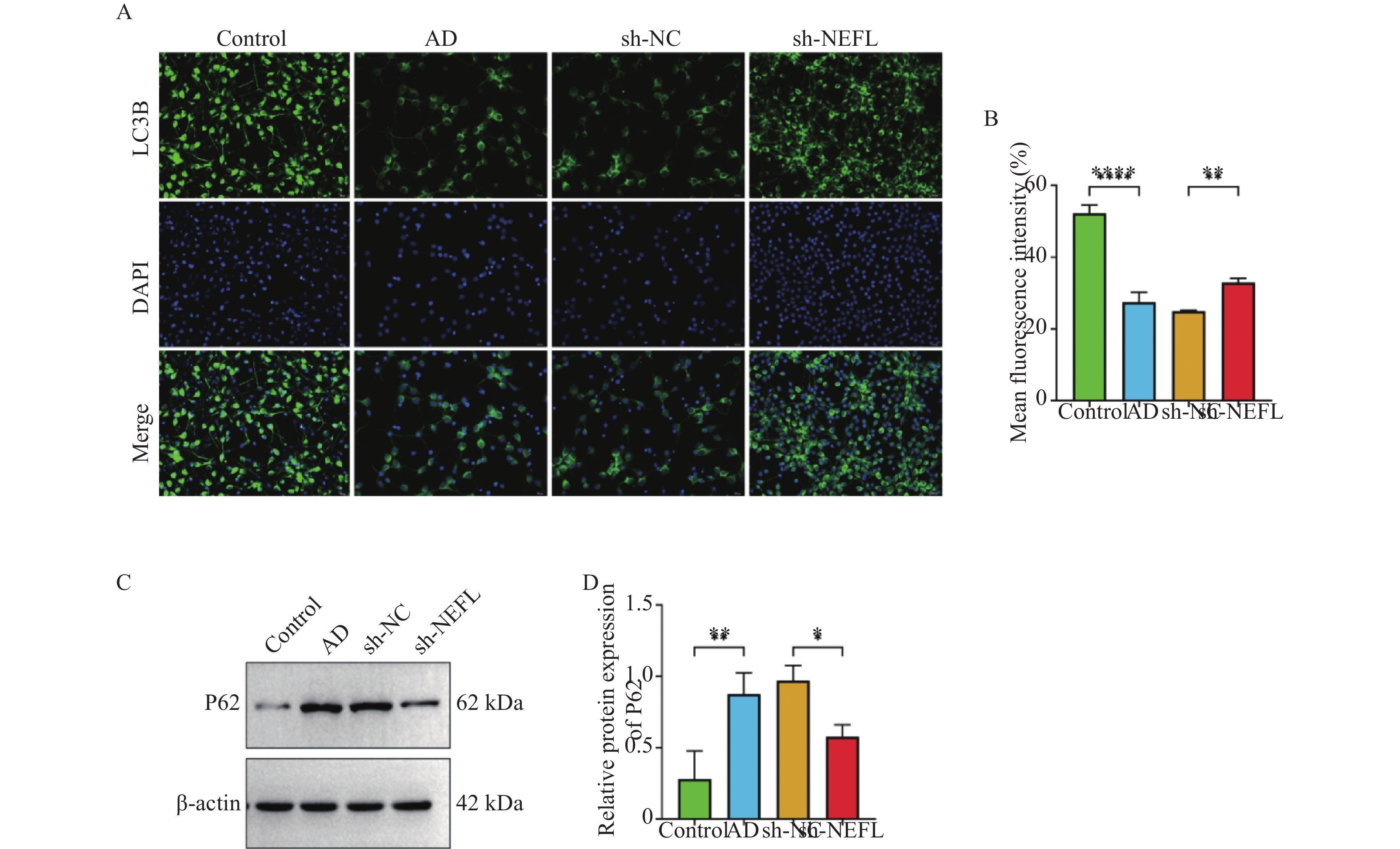
图 6 敲低NEFL缓解细胞自噬缺陷
A~B:免疫荧光染色检测LC3B的表达,40×;C~D:Western blot检测p62蛋白的表达;*P < 0.05;**P < 0.01;****P < 0.0001。
Figure 6. NEFL knockdown alleviates cellular autophagy defects
表 1 引物序列
Table 1. Primers sequence
基因 引物序列(5’-3’) 引物长度(bp) NEFL F:CTTACTCCAGCTACTCGGCG
R:CGGCATGCTTCGATTTCCAGF:20
R:20TSC1 F:AACCTGTAGCACACGTCCTG
R:CGGCTTTGCCCACATATTCGF:20
R:20GAPDH F:CCATGGGGAAGGTGAAGGTC
R:AGTGATGGCATGGACTGTGGF:20
R:20 下载: 导出CSV
-
[1] Zhang J, Zhang Y, Wang J, et al. Recent advances in Alzheimer’ s disease: Mechanisms, clinical trials and new drug development strategies[J]. Signal Transduct Target Ther, 2024, 9: 211. doi: 10.1038/s41392-024-01911-3 [2] Liu E, Zhang Y, Wang J Z. Updates in Alzheimer’ s disease: From basic research to diagnosis and therapies[J]. Transl Neurodegener, 2024, 13(1): 45. doi: 10.1186/s40035-024-00432-x [3] Zheng Q, Wang X. Alzheimer’ s disease: Insights into pathology, molecular mechanisms, and therapy[J]. Protein Cell, 2025, 16(2): 83-120. doi: 10.1093/procel/pwae026 [4] Liu Y, He Q, Zhu L, et al. TRIM29 promotes glioblastoma progression via ubiquitinating NEFL and activating the PI3K/AKT signaling pathway[J]. Cancer Genet, 2025, 296: 88-99. doi: 10.1016/j.cancergen.2025.06.008 [5] Li S, Xiao J, Huang C, et al. Identification and validation of oxidative stress and immune-related hub genes in Alzheimer’ s disease through bioinformatics analysis[J]. Sci Rep, 2023, 13: 657. doi: 10.1038/s41598-023-27977-7 [6] Huang Z, Zhuo Y, Shen Z, et al. The role of NEFL in cell growth and invasion in head and neck squamous cell carcinoma cell lines[J]. J Oral Pathol Med, 2014, 43(3): 191-198. doi: 10.1111/jop.12109 [7] Zhu S, Su L, Zhuang M, et al. NEFL modulates NRN1-mediated mitochondrial pathway to promote diacetylmorphine-induced neuronal apoptosis[J]. Mol Neurobiol, 2025, 62(6): 6983-6997. doi: 10.1007/s12035-024-04629-z [8] Mallela K, Kumar A. Role of TSC1 in physiology and diseases[J]. Mol Cell Biochem, 2021, 476(6): 2269-2282. doi: 10.1007/s11010-021-04088-3 [9] Guo Y, Chekaluk Y, Zhang J, et al. TSC1 involvement in bladder cancer: Diverse effects and therapeutic implications[J]. J Pathol, 2013, 230(1): 17-27. doi: 10.1002/path.4176 [10] Lee D F, Kuo H P, Chen C T, et al. IKKβ suppression of TSC1 links inflammation and tumor angiogenesis via the mTOR pathway[J]. Cell, 2007, 130(3): 440-455. doi: 10.1016/j.cell.2007.05.058 [11] Malhowski A J, Hira H, Bashiruddin S, et al. Smooth muscle protein-22-mediated deletion of Tsc1 results in cardiac hypertrophy that is mTORC1-mediated and reversed by rapamycin[J]. Hum Mol Genet, 2011, 20(7): 1290-1305. doi: 10.1093/hmg/ddq570 [12] Song L, Su M, Wang S, et al. miR-451 is decreased in hypertrophic cardiomyopathy and regulates autophagy by targeting TSC1[J]. J Cell Mol Med, 2014, 18(11): 2266-2274. doi: 10.1111/jcmm.12380 [13] Karalis V, Wood D, Teaney N A, et al. The role of TSC1 and TSC2 proteins in neuronal axons[J]. Mol Psychiatry, 2024, 29(4): 1165-1178. doi: 10.1038/s41380-023-02402-7 [14] 田娜, 朱敏. 阿尔茨海默病发病机制研究进展[J]. 中国实用内科杂志, 2025, 45(10): 817-822. [15] 李想, 梁文野, 蒋在军. 阿尔茨海默病治疗的研究进展[J]. 中国研究型医院, 2024, 11(4): 72-76. [16] Arzouni N, Matloff W, Zhao L, et al. Identification of dysregulated genes for late-onset Alzheimer's disease using gene expression data in brain [J]. J Alzheimers Dis Parkinsonism, 2020, 10(6). [17] Jiao X, Lu Y, Huang Y, et al. Plasma proteomic profiling reveals Parkinson’ s disease-associated proteins: A UK Biobank study[J]. Park Relat Disord, 2025, 135: 107851. doi: 10.1016/j.parkreldis.2025.107851 [18] Davoody S, Asgari Taei A, Khodabakhsh P, et al. mTOR signaling and Alzheimer’ s disease: What we know and where we are?[J]. CNS Neurosci Ther, 2024, 30(4): e14463. doi: 10.1111/cns.14463 [19] Adriaanse B A, Brady S, Wang M, et al. Tuberous sclerosis complex-1 (TSC1) contributes to selective neuronal vulnerability in Alzheimer’ s disease[J]. Neuropathol Appl Neurobiol, 2023, 49(3): e12904. doi: 10.1111/nan.12904 [20] Oddo S, Lanza M, Casili G, et al. The role of S6K1 in aging and Alzheimer’ s disease: Mechanistic insights and therapeutic potential[J]. Int J Mol Sci, 2025, 26(13): 5923. doi: 10.3390/ijms26135923 [21] Firdous S M, Ali Khan S, Maity A. Oxidative stress–mediated neuroinflammation in Alzheimer’ s disease[J]. Naunyn Schmiedeberg’ s Arch Pharmacol, 2024, 397(11): 8189-8209. doi: 10.1007/s00210-024-03188-3 [22] Heneka M T, Carson M J, El Khoury J, et al. Neuroinflammation in Alzheimer’ s disease[J]. Lancet Neurol, 2015, 14(4): 388-405. -

 点击查看大图
计量
- 文章访问数: 42
- HTML全文浏览量: 26
- PDF下载量: 33
- 被引次数: 0
