

Correlation between Epithelial Cell Related Genes and Prognosis of Patients with Ovarian Cancer based on Single Cell Sequencing
-
摘要:
目的 基于上皮细胞标志物的表达构建1个多基因风险评分来评估卵巢癌患者的预后。 方法 对卵巢癌单细胞测序数据进行降维、聚类,识别上皮细胞标记物、恶性和非恶性标记物。使用回归分析筛选与预后相关的上皮细胞标记基因以构建风险评分模型,基于风险评分将患者分为高、低风险(H.Risk、L.Risk)组,用于预测卵巢癌患者的预后。 结果 构建了1个4个基因(EPCAM、CLDN4、CXCR4和TIMP3)的风险评分模型。生存分析表明在试验队列和验证队列中H.Risk组患者的OS均比L.Risk组患者差(P < 0.05)。途径富集分析显示,高、低风险组之间的差异基因与免疫抑制和恶性进展相关,包括细胞粘附、细胞外基质、神经活性配体-受体相互作用、钙信号通路、转化生长因子-β等。 结论 通过bulkRNA-seq和scRNA-seq数据的综合分析提出了1种基于上皮细胞亚群标记基因的风险评分模型,并可能为卵巢癌患者提供潜在的治疗靶点。 Abstract:Objective To construct a polygenic risk score based on the expression of epithelial cell markers to evaluate the prognosis of patients with ovarian cancer. Methods The single cell sequencing data of ovarian cancer were reduced and clustered to identify epithelial cell markers, malignant and non-malignant markers. Regression analysis was used to screen epithelial marker genes related to prognosis to construct a risk score model. Based on the risk score, patients were divided into high risk group and low risk group (H.Risk, L.Risk) to predict the prognosis of patients with ovarian cancer. Results A risk scoring model with four genes (EPCAM, CLDN4, CXCR4 and TIMP3) was constructed. Survival analysis showed that the OS of patients in H.Risk group was worse than that in L.Risk group in trial cohort and verification cohort (P < 0.05). Pathway enrichment analysis showed that the differential genes between high and low risk groups were associated with immunosuppression and malignant progression, including cell adhesion, extracellular matrix, neuroactive ligand-receptor interaction, calcium signal pathway, transforming growth factor-β. Conclusion Through the comprehensive analysis of bulkRNA-seq and scRNA-seq data, a risk scoring model based on epithelial cell subsets marker genes is proposed, which may provide potential therapeutic targets for patients with ovarian cancer. -
Key words:
- Ovarian cancer /
- Epithelial cells /
- Single cell RNA sequencing /
- Bioinformatics /
- Prognosis
-
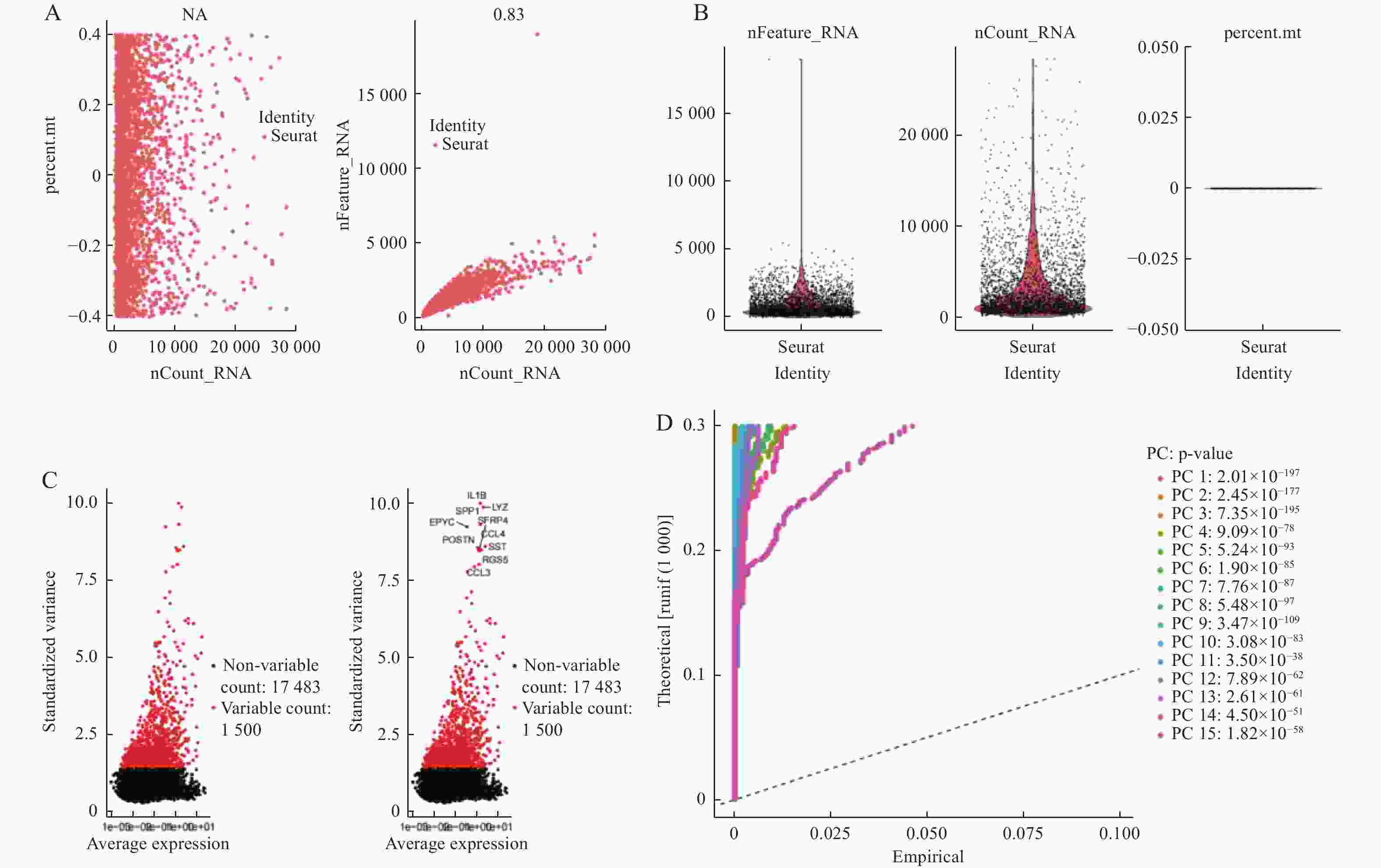
图 1 卵巢癌的单细胞RNA测序分析
A:线粒体基因与测序深度的关系(线粒体基因含量为0)和测序深度与基因数目呈正相关关系;B:线粒体基因含量;C:前1500个高变基因;D:前15个主成分。
Figure 1. Single cell RNA sequencing analysis of ovarian cancer
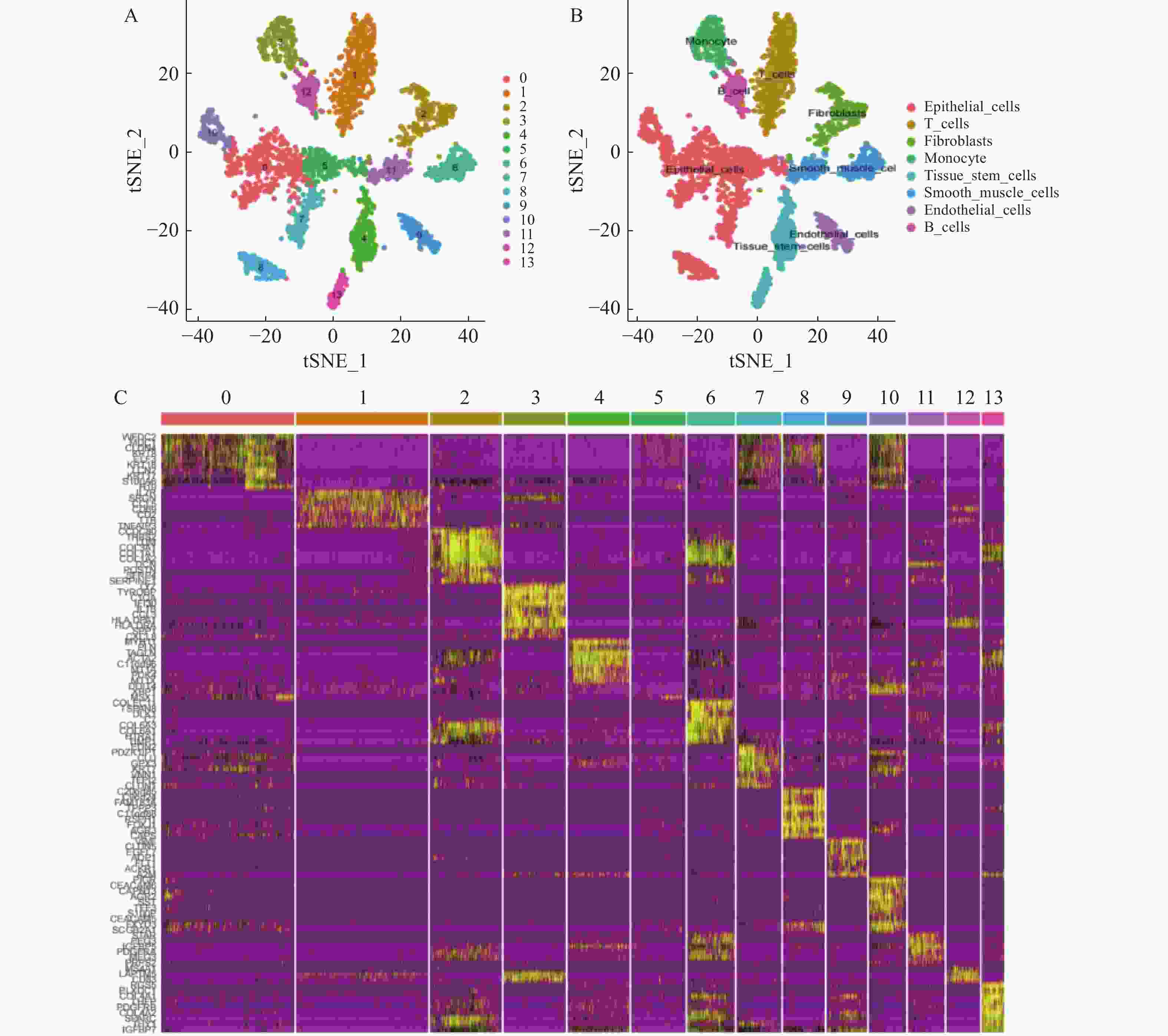
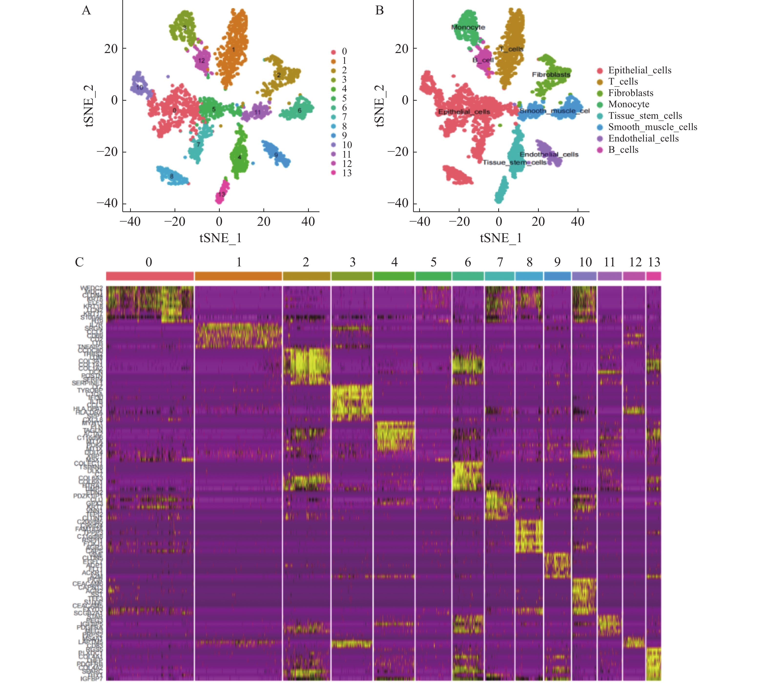
图 2 单细胞数据的降维和聚类
A:细胞的T-SNE图,显示细胞群;B:细胞注释的T-SNE图;C:细胞群标记物的相对表达热图(仅显示前10名)。
Figure 2. Dimensionality reduction and clustering of single cell data
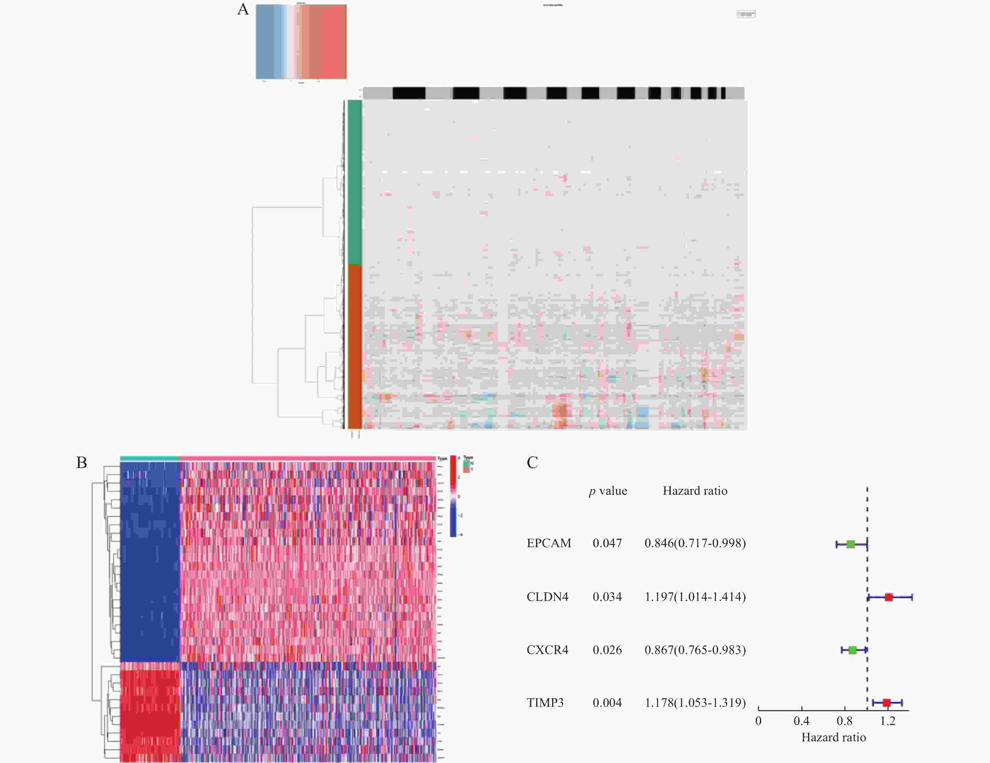
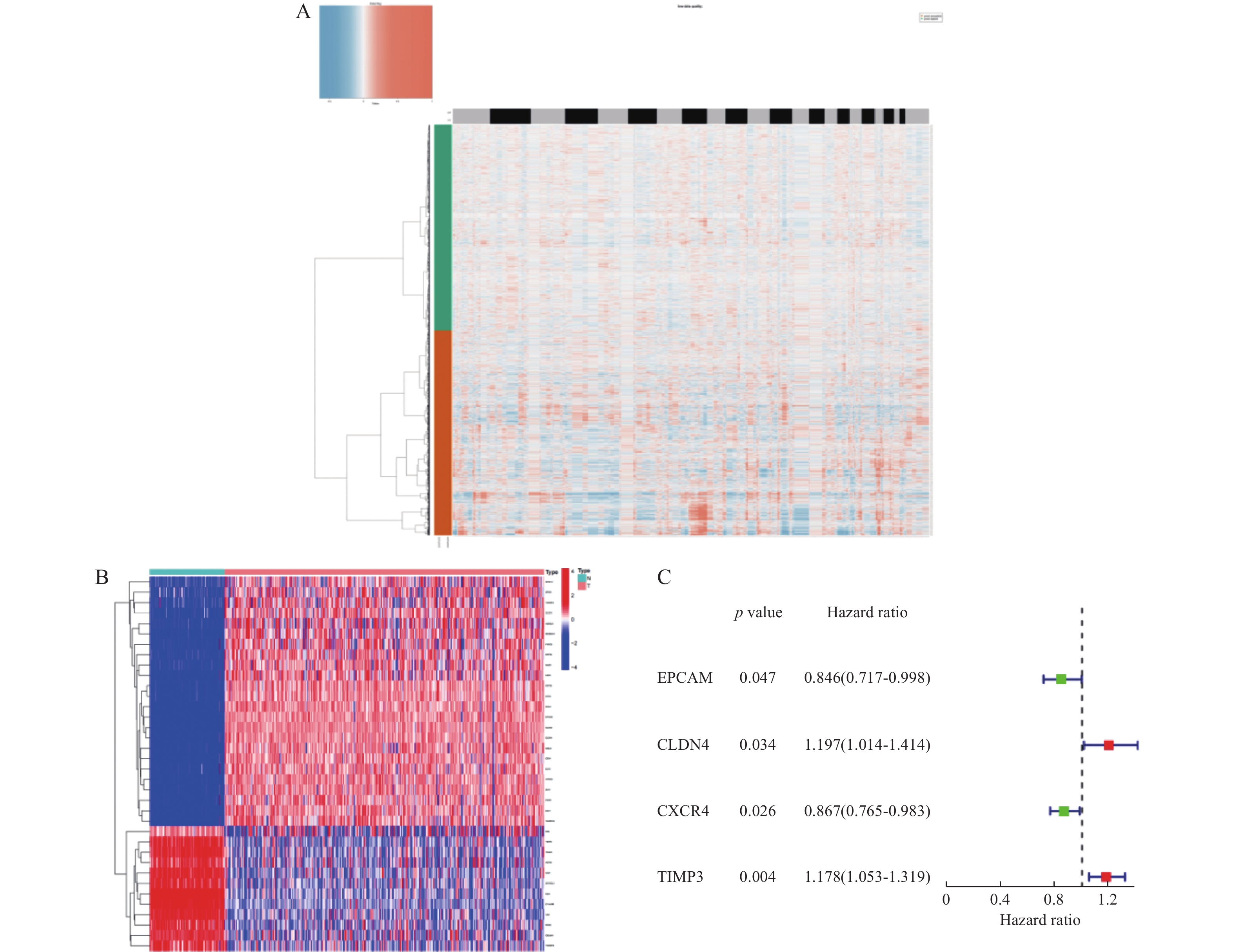
图 3 单细胞CNV分析及预后基因筛选
A:拷贝组变异分析热图;B:MECRGs在良恶性患者中的表达热图;C:基因表达和OS之间单变量Cox回归分析结果的森林图。
Figure 3. Single cell CNV analysis and prognosis gene screening
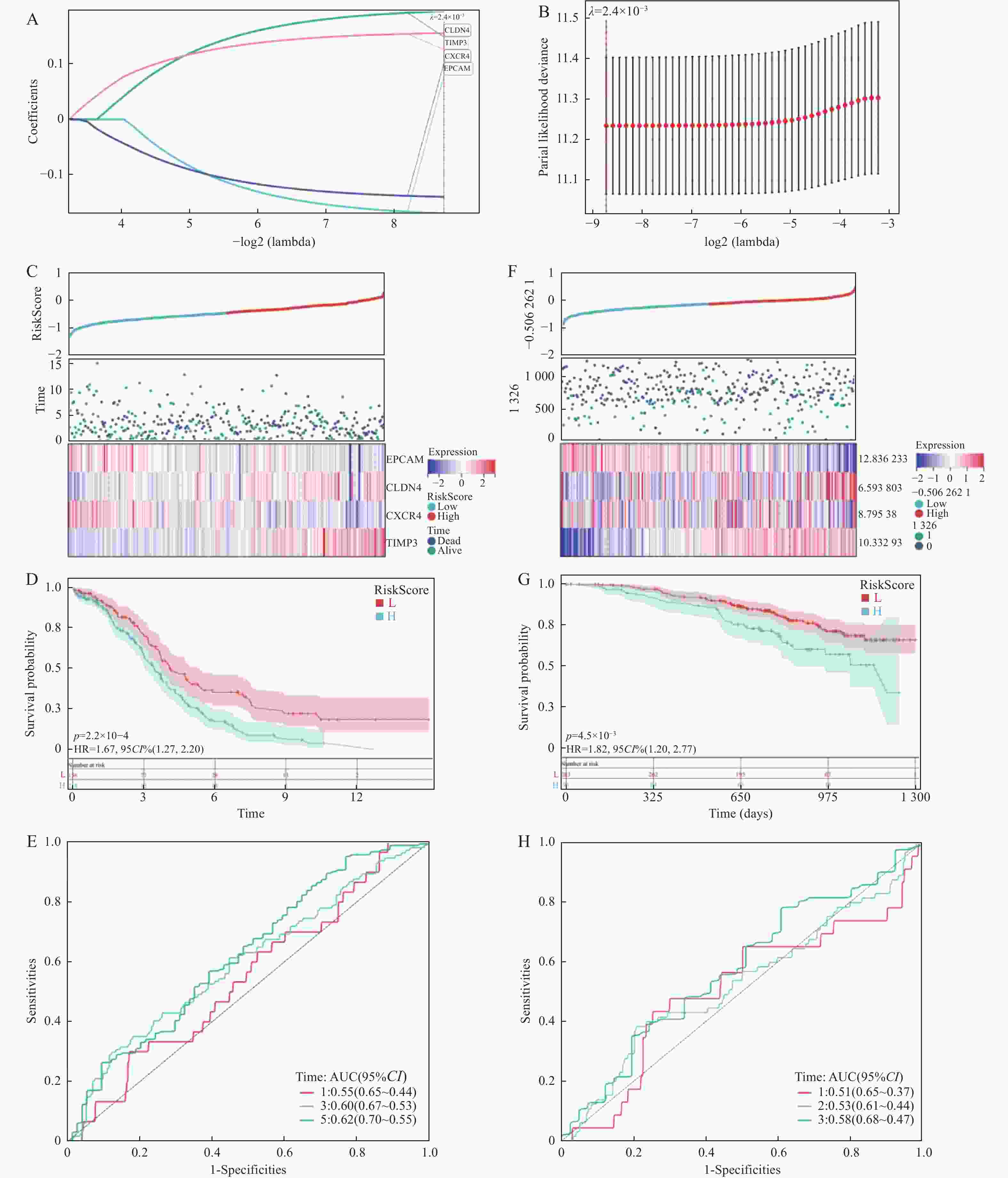
图 4 TCGA-OV中的多基因风险评分构建与验证
A~B:Lasso-cox回归分析;C:TCGA队列中OS状态、OS和风险评分的分布、模型基因的表达热图;D:TCGA-OV队列高、低风险组患者OS的K-M曲线(P < 0.001);E:TCGA-OV队列ROC曲线;F:GEO队列中OS状态、OS和风险评分的分布、模型基因的表达热图;G:GEO队列高低风险组患者OS的K-M曲线(P < 0.001);H:GEO队列ROC曲线。
Figure 4. Construction and verification of polygene risk score in 4TCGA-OV
-
[1] Menon U,Karpinskyj C,Gentry-Maharaj A. Ovarian cancer prevention and screening[J]. Obstetrics & Gynecology,2018,131(5):909-927. [2] Jemal A,Siegel R,Ward E,et al. Cancer statistics,2008[J]. CA:A Cancer Journal for Clinicians,2008,58(2):71-96. doi: 10.3322/CA.2007.0010 [3] Karantza V. Keratins in health and cancer: More than mere epithelial cell markers[J]. Oncogene,2011,30(2):127-138. doi: 10.1038/onc.2010.456 [4] Tanimura N, Fujita Y. Epithelial defense against cancer (EDAC)[C]. Seminars in Cancer Biology, 2020, 63(6): 44-48. [5] Royer C,Lu X. Epithelial cell polarity: A major gatekeeper against cancer?[J]. Cell Death & Differentiation,2011,18(9):1470-1477. [6] Bai Z,Woodhouse S,Zhao Z,et al. Single-cell antigen-specific landscape of CAR T infusion product identifies determinants of CD19-positive relapse in patients with ALL[J]. Science Advances,2022,8(23):eabj2820. doi: 10.1126/sciadv.abj2820 [7] Parker K R,Migliorini D,Perkey E,et al. Single-cell analyses identify brain mural cells expressing CD19 as potential off-tumor targets for CAR-T immunotherapies[J]. Cell,2020,183(1):126-142.e17. doi: 10.1016/j.cell.2020.08.022 [8] Varga J,Greten F R. Cell plasticity in epithelial homeostasis and tumorigenesis[J]. Nature Cell Biology,2017,19(10):1133-1141. doi: 10.1038/ncb3611 [9] Chen Z,Zhang H,Bai Y,et al. Single cell transcriptomic analysis identifies novel vascular smooth muscle subsets under high hydrostatic pressure[J]. Science China Life Sciences,2021,64(1):1677-1690. [10] Pan J,Zhou H,Cooper L,et al. LAYN is a prognostic biomarker and correlated with immune infiltrates in gastric and colon cancers[J]. Frontiers in Immunology,2019,10(1):6. [11] Lombardo G,Gili M,Grange C,et al. IL-3R-alpha blockade inhibits tumor endothelial cell-derived extracellular vesicle (EV)-mediated vessel formation by targeting the β-catenin pathway[J]. Oncogene,2018,37(9):1175-1191. doi: 10.1038/s41388-017-0034-x [12] Ichimiya H,Maeda K,Enomoto A,et al. Girdin/GIV regulates transendothelial permeability by controlling VE-cadherin trafficking through the small GTPase,R-Ras[J]. Biochemical and Biophysical Research Vommunications,2015,461(2):260-267. doi: 10.1016/j.bbrc.2015.04.012 [13] Gires O,Pan M,Schinke H,et al. Expression and function of epithelial cell adhesion molecule EpCAM: Where are we after 40 years?[J]. Cancer and Metastasis Reviews,2020,39(6):969-987. [14] Corso G,Figueiredo J,De Angelis S P,et al. E‐cadherin deregulation in breast cancer[J]. Journal of Cellular and Molecular Medicine,2020,24(11):5930-5936. doi: 10.1111/jcmm.15140 [15] Fang L,Yu G,Yu W,et al. The correlation of WDR76 expression with survival outcomes and immune infiltrates in lung adenocarcinoma[J]. Peer J,2021,9(10):12277. [16] Liu Y,Wang Y,Sun S,et al. Understanding the versatile roles and applications of EpCAM in cancers: From bench to bedside[J]. Experimental Hematology & Oncology,2022,11(1):1-19. [17] Yahyazadeh Mashhadi S M,Kazemimanesh M,Arashkia A,et al. Shedding light on the EpCAM: An overview[J]. Journal of Cellular Physiology,2019,234(8):12569-12580. doi: 10.1002/jcp.28132 [18] Driemel C,Kremling H,Schumacher S,et al. Context-dependent adaption of EpCAM expression in early systemic esophageal cancer[J]. Oncogene,2014,33(41):4904-4915. [19] Yoon S M,Gerasimidou D,Kuwahara R,et al. Epithelial cell adhesion molecule (EpCAM) marks hepatocytes newly derived from stem/progenitor cells in humans[J]. Hepatology,2011,53(3):964-973. [20] Uthayanan L,El-Bahrawy M. Potential roles of claudin-3 and claudin-4 in ovarian cancer management[J]. Journal of the Egyptian National Cancer Institute,2022,34(1):1-9. doi: 10.1186/s43046-021-00099-9 [21] Hicks D A,Galimanis C E,Webb P G,et al. Claudin-4 activity in ovarian tumor cell apoptosis resistance and migration[J]. BMC Cancer,2016,16(1):1-11. doi: 10.1186/s12885-015-2026-y [22] Yamamoto T M,Webb P G,Davis D M,et al. Loss of claudin-4 reduces DNA damage repair and increases sensitivity to PARP inhibitors[J]. Molecular Cancer Therapeutics,2022,21(4):647-657. [23] English D P,Santin A D. Claudins overexpression in ovarian cancer: potential targets for Clostridium Perfringens Enterotoxin (CPE) based diagnosis and therapy[J]. International Journal of Molecular Sciences,2013,14(5):10412-10437. [24] Jacobson O,Weiss I D. CXCR4 chemokine receptor overview: Biology,pathology and applications in imaging and therapy[J]. Theranostics,2013,3(1):1. doi: 10.7150/thno.5760 [25] Zhou Y,Zhang T,Wang S,et al. Targeting of HBP1/TIMP3 axis as a novel strategy against breast cancer[J]. Pharmacological Research,2023,194(8):106846. [26] Pol M,Gao H,Zhang H,et al. Dynamic modulation of matrix adhesiveness induces epithelial-to-mesenchymal transition in prostate cancer cells in 3D[J]. Biomaterials,2023,299(16):122180. [27] 陈鑫磊,余永波,李鹏,等. 转化生长因子β1对膀胱癌细胞增殖与迁移能力的影响及其机制[J]. 精准医学杂志,2023,38(2):111-115. -

 下载:
下载:


 点击查看大图

计量
- 文章访问数: 645
- HTML全文浏览量: 425
- PDF下载量: 26
- 被引次数: 0
